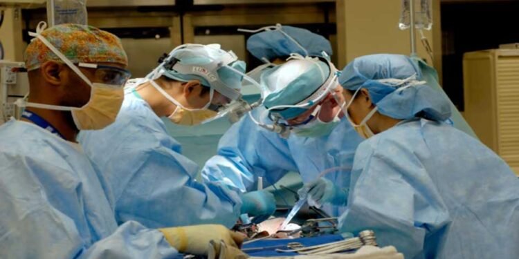
Nerve damage during amputation surgery can have devastating consequences for patients, severely impacting their quality of life and ability to use prosthetic limbs. As surgeons, preserving nerve function should be one of your top priorities whenever performing amputations. Fortunately, there are several minimally invasive techniques that may help reduce the risk of nerve injury. This article will discuss 5 techniques that promise to enhance nerve preservation outcomes.
5 Ways To Protect Nerves in Amputation Surgery
As amputation surgeons, adopting a nerve-first approach and using minimally invasive techniques can significantly impact patients’ quality of life long after surgery. Here are 5 key ways to prioritize nerve preservation when performing amputations:
#1 Intraoperative Neuromonitoring (IONM)
IONM involves using electrical stimulation and monitoring equipment during surgery to locate and map nerves in real time. This allows surgeons to visualize nerve pathways that may otherwise be difficult to identify, especially in cases with extensive scar tissue or previous injuries.
For amputations, IONM helps ensure nerves are not inadvertently cut or damaged. Surgeons can adjust their technique based on the nerve monitoring feedback to safely resect tissues in close proximity to nerves.
IONM has been shown to significantly reduce postoperative neuroma formation and neuropathic pain levels compared to amputations performed without monitoring.
#2 Nerve Blocks and Topical Anesthetics
Preserving nerve function also means minimizing unnecessary trauma to nerves during and after surgery. Providing effective pain management through nerve blocks and topical anesthetics can help reduce intraoperative and postoperative neuroma formation.
For example, administering a sciatic nerve block before a right-sided transfemoral amputation numbs the leg and helps the surgeon gently dissect tissues near the sciatic nerve without causing additional nerve injury from surgical handling or traction.
Topical anesthetics applied to severed nerve ends may also reduce neuroma formation by dampening the nerve impulse traffic in the initial postoperative period.
#3 Microsurgical Techniques
In some cases where major nerves like the sciatic or femoral nerve need to be resected or repaired, microsurgical techniques can optimize outcomes.
Using an operating microscope, microsurgical instruments, and fine sutures allows for the most precise reconstruction of severed nerve ends with the aim of restoring nerve conduction.
While microsurgery requires additional training, it may be worthwhile considering for young, active patients undergoing amputations where maximizing return of function is a high priority. Several studies have found that microsurgical nerve repairs can result in more recovery of motor and sensory function after amputation.
#4 Nerve Grafting and Nerve Transfers
When a nerve defect is too large to directly repair, alternative reconstructive options like nerve grafts or nerve transfers may be considered. Nerve grafts involve taking a donor nerve, such as a sural nerve, and using it to bridge the gap between the proximal and distal nerve stumps.
Nerve transfers are also gaining popularity, such as transferring part of an expendable nerve, like the nerve to the tibialis anterior muscle to repair a damaged sciatic nerve.
Both grafts and transfers can reinnervate distal tissues and restore some level of sensory or motor function, preventing permanent denervation if primary repair is impossible.
#5 Neuroma Management
Even with the most meticulous surgical techniques, neuromas may still form at amputation sites. Two minimally invasive options for neuroma management include capping the nerve end with local tissues or a nerve conduit. This physical barrier prevents the neuroma from impinging on surrounding tissues and causing pain.
Neuroma injections with corticosteroids or other agents can also help reduce pain and swelling levels. Combined with a compression sleeve or shoe, these conservative measures may control neuroma pain sufficiently without the need for reoperation in numerous instances.
Conclusion
Amputation surgery challenges nerve preservation, but adopting minimally invasive techniques like IONM, careful dissection, microsurgery, nerve reconstruction, and neuroma management can significantly improve postoperative outcomes. With diligent nerve protection strategies, surgeons can maximize remaining function and quality of life for patients undergoing lower extremity amputations.


